Epifluorescence Microscopy
The platform has two widefield microscopes for the acquisition of material fixed in fluorescence, a widefield microscope equipped with a colour camera, a routine fluorescence microscope for the observation of living cells and an analysis computer equipped with Fiji and Icy software. This equipment is reserved for the internal use of UMR7216 members.

Artistic image of jellyfish, referring to the bioluminescent jellyfish from which GFP (Green Fluorescent Protein) is extracted that has revolutionised the world of imaging.
© Image par Carol-Ann Bussières de Pixabay
Leica DMI-6000B "Oldelaf"
Inverted epifluorescence microscope
X, Y and Z piloted
Filters and objectives piloted
Camera:
Camera CCD -30ºC regulated
CoolSNAP HQ2 14bits
Image size: 1392×1040 pixels
Pixel size: 6.45 x 6.45μm
Illumination:
Visible light (halogen)
CoolLED pE-300 White Series Fluorescent Lamp (LED, >25000h)
Acquisition software:
Metamorph with Multi-Dimensional Acquisition module
Objectives:
| Obj Mag. | N.A | Quality | Transmission Contrast | Imm. | Pixel size (binning 1) | Max resolution at 488nm |
| 10X | 0,25 | N PLAN | PH1 | Air | 0,645 μm | 1191 nm |
| 20X | 0,35 | N PLAN | PH1 | Air | 0,3225 μm | 850 nm |
| 40X | 0,6 | HCX PL Fluotar | PH2 | Air | 0,161 μm | 496 nm |
| 40X | 1,25-0,75 | HC PL APO CS | – | Oil | 0,161 μm | 229 nm |
| 63X | 1,40 – 0.60 | HCX PL APO | – | Oil | 0,102 μm | 213 nm |
| 100X | 1,40 | HCL PL APO CS | PH3 | Oil | 0,0645 μm | 213 nm |
Filters :
| Filter name | Colour | Excitation Filter BP | Dichroic Mirror | Suppression Filter BP |
| A4 | Blue | 360/40 | 400 | 470/40 |
| GFP | Green | 470/40 | 500 | 525/50 |
| Y3 | Cy3 (orange) | 545/40 | 565 | 610/75 |
| TX2 | Red | 560/40 | 595 | 645/75 |
| Y5 | Cy5 (Far red) | 620/60 | 660 | 700/75 |
| Analyzer cube | – | – | – | Analyzer |
Leica DMI-6000B "Lenny"
Inverted epifluorescence microscope
Controlled in X, Y and Z
Controlled filters and lenses
Camera:
Camera CCD -12ºC regulated
Teledyne Photometrics RETIGA R6
Image size: 2688×2200 pixels
Pixel size: 4.54 x 4.54μm
Illumination:
Visible light (halogen)
CoolLED pE-300 White Series Fluorescent Lamp (LED, >25000h)
Acquisition software:
Metamorph with Multi-Dimensional Acquisition module
Objectives:
| Mag. | N.A | Quality | Trans Contrast | Imm. | Pixel size (binning 1) | Max resolution at 488 nm |
| 2,5X | 0,07 | HC FL PLAN | – | Air | 1.816 μm | 4,2 µm |
| 20X | 0,7 | HCL PL APO CORR | – | Oil/Gly | 0,227 μm | 425 nm |
| 40X | 1,30 | HC PL APO | – | Oil | 0,1135 μm | 229 nm |
| 63X | 1,40 | HCX PL APO CS | PH3 | Oil | 0,072 μm | 212 nm |
| 100X | 1,40-0,7 | HCX PL APO | – | Oil | 0,0454 μm | 212 nm |
Filters :
| Name | Colour | Excitation Filter BP | Dichroic Mirror | Suppression Filter BP |
| XF131 | Blue | 387/28 | 410 | 450/65 |
| QMAX green | Green | 450-490 | 500 | 510-560 |
| N3 | Cy3 (orange) | 546/12 | 565 | 600/40 |
| TX2 | Red | 560/40 | 595 | 645/75 |
| Far Red | Cy5 | 620-60 | 660 | 700-75 |
| Analyzer cube | – | – | – | Analyzer |
Leica DM IL LED (Live)
Inverted epifluorescence microscope
Camera:
Camera CCD -60ºC regulated
Hamamatsu digital camera C4742-98-24ERG 12 or 14 bits
Image size: 1344×1024
Pixel size: 6.45 x 6.45μm
Illumination:
Visible light (halogen)
Leica fluorescent lamp (Mercury Halides – 3000h max)
Objectives:
| Mag. | N.A | Quality | Transmission Contrast |
Imm. | Pixel size (binning 1) |
Max resolution at 488 nm |
| 10X | 0,25 | N PLAN | PH1 | Air | 0,645 μm | 1,2 µm |
| 20X | 0,35 | N PLAN | PH1 | Air | 0,322 μm | 850 nm |
| 40X | 0,55 | CORR | PH2 | Air | 0,161 μm | 541 nm |
Filtres :
| Name | Colour | Excitation Filter BP | Dichroic Mirror | Suppression Filter BP |
| B/G/R | Blue/Green/Red | 420/30 ; 495 /15 ; 570/20 | 415 ; 510 ; 590 | 465/20 ; 530 /30 ; 640/40 |
| GFP ET | Green | |||
| TX ET | Red |
Leica DMRA2 (Color)
Upright epifluorescence microscope
Controlled in X, Y and Z
Camera:
CCD digital color camera 5Mpixels Leica DFC 450C cooled (Δ -20ºC compared to ambient)
Image size: 2560×1920
Pixel size: 3.4 x 3.4μm
Illumination:
Visible light (halogen)
Fluorescent lamp not installed
Objectives:
| Mag. | N.A | Quality | Transmission Contrast |
Imm. | Pixel size (binning 1) |
Max resolution at 488 nm |
| 10X | 0,30 | HC PL Fluotar | – | Air | 0,34 μm | 992 nm |
| 20X | 0,50 | HCX PL Fluotar | – | Air | 0,17 μm | 595 nm |
| 40X | 0,75 | HCX PL Fluotar | – | Air | 0,085 μm | 397 nm |
| 100X | 1,40-0,7 | HCX PL APO CS | – | Oil | 0,034 μm | 212 nm |
Workstation "Angus"
A workstation for image analysis is available. This workstation is equipped with the free software FIJI, IMAGEJ, ICY.
Read more

Congratulations to the Defossez group for their review published in Médecine/Sciences
Congratulations to Matthieu, Raady and Pierre-antoine for their review "Demethylating DNA against cancer : towards new epigenetic therapeutic strategies without DNA damage" published in Médecine/Sciences in february 2026! Read here
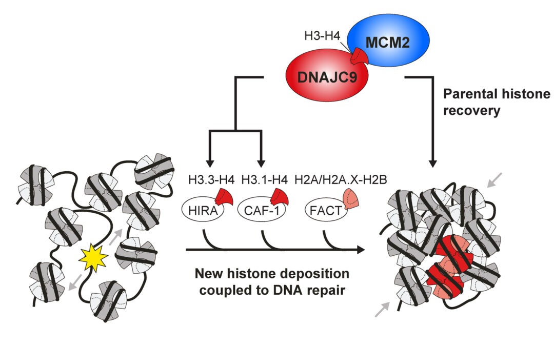
New research article on histone chaperones with central functions in chromatin repair
In this paper, we identify new players in chromatin repair through a novel approach for the proteomic profiling of DNA repair patches in human cells. Thus, we expand the histone chaperone network involved in repair-coupled chromatin restoration and highlight a...

Welcome to Léa
Léa joins the team as a research assistant. After completing a master's degree in virology, she worked in Strasbourg on grapevine viruses, then on characterizing mRNA degradation in plants at the Institute of Plant Molecular Biology (IBMP). In the Polo team, Léa will...

Sophie Polo receives an Impulscience® grant from the Fondation Bettencourt Schueller
Sophie Polo has been awarded an Impulscience® grant to fund a research project on the establishment and maintenance of the inactive X chromosome in response to DNA breaks. This is wonderful news for the lab ! We thank the Fondation Bettencourt Schueller for their...